This coloured scanning electron microscope image shows the surface of a kidney stone (Image by Kempf EK/ Wikimedia Commons)
Question from Jacob Crowley
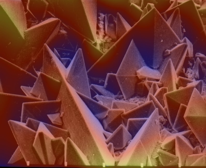
Electron microscopes can view things in much more detail than light-based microscopes. To do this they use a beam of high-energy electrons to scan the surface of the specimen placed under the microscope. When the electrons hit the specimen, they are reflected and directed towards a detector, creating an image of the sample.
Because the electrons’ wavelengths are much shorter than that of light, the image is much more detailed and can show small specimens such as bacteria or the minute details of metals and crystals.
To feature in our Brain Dump section, send us your questions tohowitworks@futurenet.comor message us on Instagram,Facebook or TwitterFor more science and technology articles, pick up the latest copy of How It Works from all good retailers or from ourwebsitenow. If you have a tablet or smartphone, you can also download the digital version onto youriOSorAndroiddevice. To make sure you never miss an issue of How It Works magazine,subscribe today!