How does a CT scanner work?
by Scott Dutfield · 22/03/2019
These 3D X-rays can create detailed pictures of the inside of your body
At the end of the 19th century, Wilhelm Röntgen discovered X-rays and changed medicine forever. As X-rays pass through the body, different tissues absorb different amounts of energy, leaving shadows on photographic film. For the first time, doctors could see inside their patients without having to cut them open. But the story didn’t stop there.
If you capture one X-ray image you see a snapshot of the body, but with the organs piled on top of one another it’s hard to make out what’s going on. In 1972, Godfrey Hounsfield found a solution when he invented computerised tomography (CT) scans, thereby revolutionising medicine again.
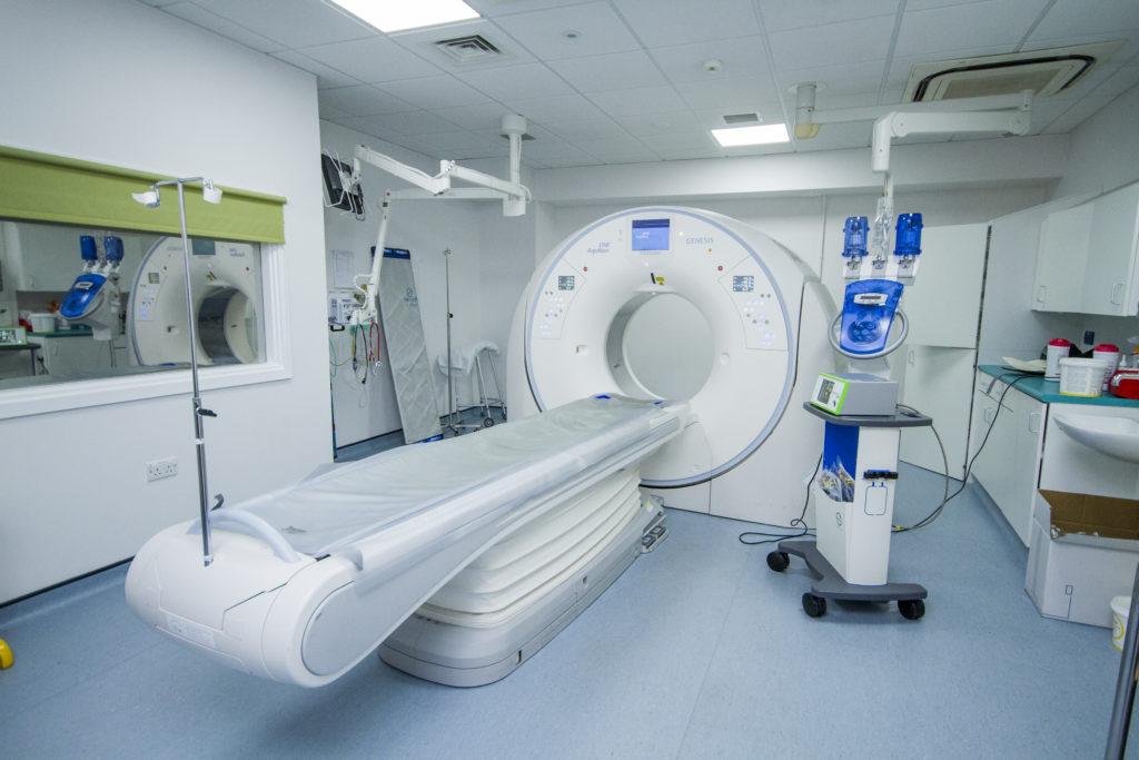
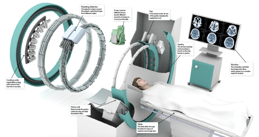
CT scanners use a rotating ring to take X-ray images from all angles. A computer then combines these images to separate out the signals from different bones, organs and blood vessels. This creates image slices between one and ten millimetres thick, showing the inside of the body in cross-section. During the scan, a table slides the patient through the ring, capturing more and more image slices. Then the computer stacks them together to make 3D pictures of the internal organs.
The result is a much higher-resolution picture of the inside of the body. The outlines of the tissues are clearer than a normal X-ray, and the 3D shapes allow medical professionals to see abnormalities. X-ray-absorbing chemicals called contrast agents can make the pictures even clearer. For example, iodine injected into the blood can reveal the outline of the blood vessels, showing up clots. Barium swallowed in a meal or drink can highlight the outline of the digestive system, revealing tumours.
Though X-rays do deliver small amounts of ionising radiation, which can damage cells, the benefits far outweigh the risks.
Inside a CT scanner
CT vs MRI
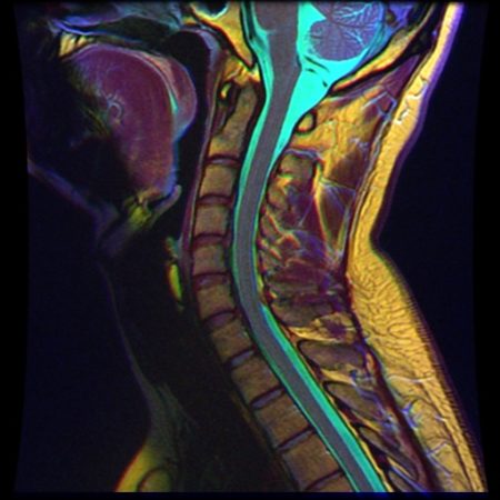
CT scans are good for showing up bones, blood vessels and organs, but they can’t capture the fine detail of soft tissues. To do this we need Magnetic Resonance Imaging (MRI). These scans use a combination of radio waves and powerful magnets to make 3D pictures. The magnets pull on the hydrogen atoms in the water molecules inside the body, rotating them so they all point in the same direction. Radio waves then knock them temporarily out of line; when they snap back in line they release energy. Detectors pick this energy up, creating a picture of where the water molecules are. Different tissues contain different amounts of water, giving a clearer view of the internal organs.
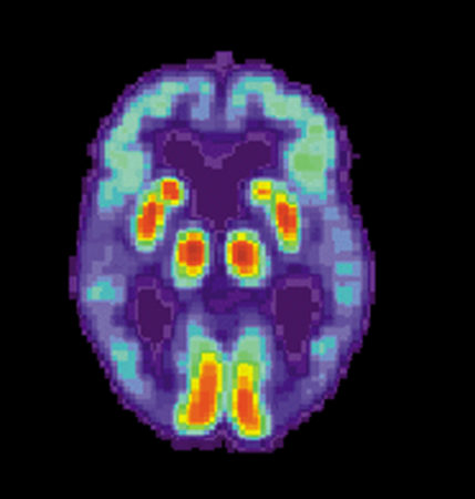
PET and SPECT
Doctors can zone in on specific parts of the body using nuclear medicine. This involves injecting, inhaling or swallowing small amounts of radioactive material to light up different tissues. Doctors sometimes need to highlight the blood vessels to look for circulation problems. To do this they can use Single Photon Emission Computed Tomography (SPECT) scans. Patients receive an injection containing radioactive atoms, which enter the blood and release gamma rays as they circulate. When a gamma camera detects the rays, it reveals the outline of the blood vessels. Another option is a Positron Emission Tomography, or PET scan. These use radioactive tracers that produce positrons instead of gamma rays. Positrons interact with electrons inside the body, sending bursts of energy to the detectors. PET tracers attached to sugar molecules can light up tissues using lots of energy, like active areas of the brain or growing tumours.
© Nevit Dilmen
This article was originally published in How It Works issue 117, written by Laura Mears
For more science and technology articles, pick up the latest copy of How It Works from all good retailers or from our website now. If you have a tablet or smartphone, you can also download the digital version onto your iOS or Android device. To make sure you never miss an issue of How It Works magazine, subscribe today!