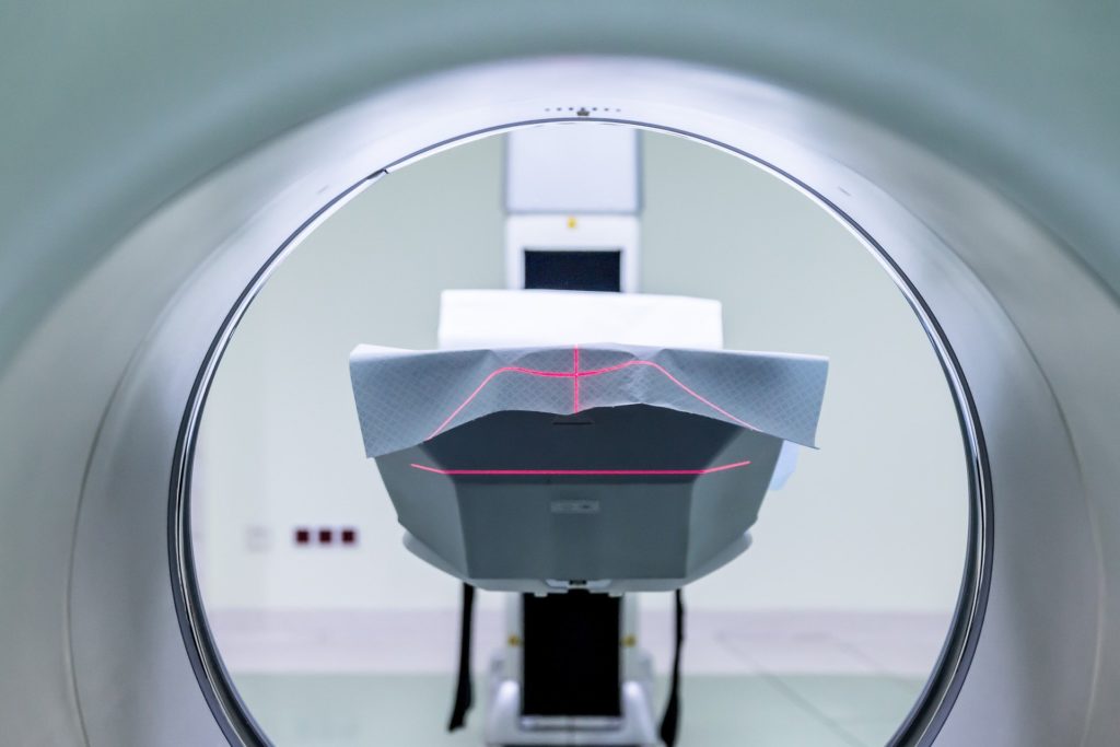
Inside an MRI scanner

When doctors need the highest quality images possible they turn to MRI scanners, but how do they work?

(Image by Michal Jarmoluk from Pixabay)

Doctors often plan treatments based on imaging. X-rays, ultrasound and CT scans provide useful pictures, but when the highest quality images are needed, they turn to MRI scanners. While CT scanners use x-rays and therefore expose the patient to radiation, magnetic resonance imaging (MRI) uses powerful magnets and is virtually risk free. 

MRI scans are obtained for many medical conditions, although since they are expensive and complicated to interpret, they certainly aren’t as easy as taking a chest x-ray. Examples for which they are used include planning surgery for rectal cancers, assessing bones for infection (osteomyelitis), looking at the bile ducts in detail for trapped gallstones, assessing ligamental damage in the knee joints and assessing the spinal cord for infections, tumours or trapped nerves.

Physicists and engineers use and manipulate the basic laws of physics to develop these incredible scanners for doctors to use. MRI scans provide such details because they work at a sub- molecular level; they work on the protons within hydrogen atoms. By changing the position of these protons using magnetic fields, extremely detailed pictures of the different types of pictures are obtained. Since these pictures rely on the tiny movements of these tiny particles, you need to lie very still during the scan.

Slice by slice images

Specially wound coils, known a gradient coils, allow for the detailed depth imaging which creates the slice by slice pictures. While the main superconducting magnet creates a very stable magnetic field, these gradient coils create variable magnetic fields during the scan. These fields mean that the magnetic strength within the patient can be altered in specific areas. Since the protons realign at different rates in different tissue types, the relationship between the strength of the field and the frequency of the emitted photons is different for various tissues.

Detecting these differences allows for very detailed images. Powerful computers outside the main machine then reconstitute all of this data to produce slice by slice imaging. Depending on what’s being scanned, 3D reconstructions can then be created, such as for brain tumours.

MRI atoms : It's a matter of reading the alignment

Line up please

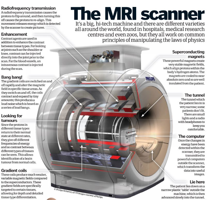
Hydrogen atoms contain just one proton and emit tiny magnetic fields. When placed in a stronger magnetic field (the one produced by the magnets), these protons line up in the direction of the field.
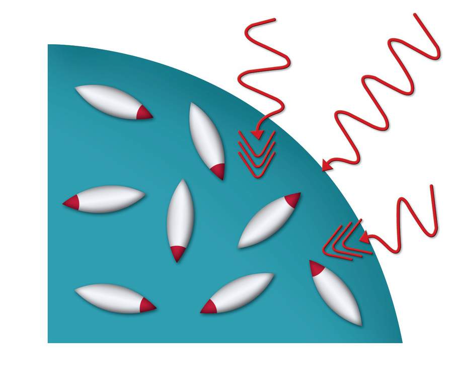
Flip and spin

The scanner emits a radiofrequency through the patient, which flips the spinning direction of these aligned protons. The frequency is at just the right pitch, producing a ‘resonance’ energy (hence magnetic resonance).
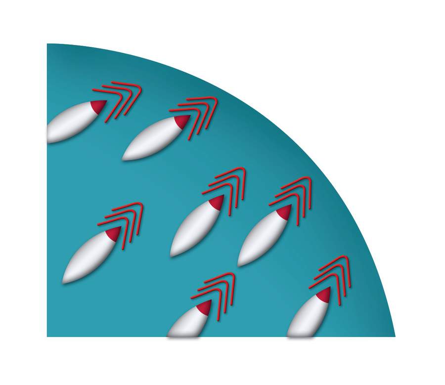
Flip back

Once the radiofrequency is removed, the protons degrade back to their original positions. As they do so, they release tiny amounts of radiowave energy in the form of photons. It is these changes that build the detailed pictures.

Converting to pictures

Different magnetic strengths produce different frequencies in the protons, which are also affected by the different type of body tissues. The resultant energy given off by re-aligning the protons is interpreted by a computer to produce detailed images.


This article was originally published in How It Works issue 13


For more science and technology articles, pick up the latest copy of How It Works from all good retailers or from our website now. If you have a tablet or smartphone, you can also download the digital version onto your iOS or Android device. To make sure you never miss an issue of How It Works magazine, subscribe today!